David Bernard, Dage Precision Industries
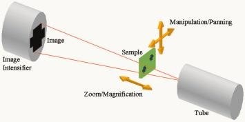
Using X-ray inspection for both board fault analysis and process control has been around for a long time. The technique relies on X-rays passing through the assembly under inspection and being partially absorbed, to a greater or lesser extent, depending on the density and other characteristics of the different materials through which the X-radiation has to go. This produces a shadow of various contrast levels at the X-ray detector; this image then represents the sample (see figure 1).
In this way, the dense solder balls in BGA packages, either lead or lead-free for example, appear black on an X-ray image, because they absorb most of the X-rays, preventing any from hitting the detector. In comparison, areas of voiding within a solder ball or other solder joints will appear as bright spots against the dark background of the joint because more X-rays hit the detector from these less dense areas. This approach is very similar to that used in hospitals for X-raying patients. The difference for inspection in electronics assembly is that higher magnification is required to see the ever-shrinking components and their details, something that is, for example, not necessary for a broken arm or leg. The magnification is achieved by moving the sample closer to the X-ray source (principle shown in figure 1), so casting a magnified shadow at the detector for capture.
In the past, X-ray inspection meant using special film material to produce the images, requiring wet chemistry expertise and equipment to then develop the film before even attempting to diagnose the fault(s). Using those films also produces a so-called negative image, compared to what we see as standard. The entire system/process of exposure, development and film properties leads to the effect that less dense material areas blacken the film (receiving more radiation), and that’s why the broken bone in an arm appears white on the X-ray. This principle follows the familiar technique of photographic negative film processing. However, in the electronics inspection and other application areas, the obsolete film-based method has generally been replaced by ‘real-time’ X-ray imaging techniques for easier handling, and the images are therefore available very quickly. This has been achieved by using X-ray devices called image intensifiers.
An image intensifier comprises of an area of X-ray sensitive phosphor that converts any incoming X-ray photons that hit the phosphor into visible light at that location. This visible light is then captured onto an analog charge-coupled device (CCD)-based camera that is looking at the phosphor target. The resulting analog image is then fed to the user of the X-ray system, and shows the variation of the received signal across the sample. Conversion of the analog data to a digital image together with image enhancement and adjustment may be necessary before presenting the image to the operator. This is achieved through some computer-based manipulation before display. The drive for image-intensifier development has come from medical requirements for real-time X-ray images during interventional radiological examinations, where the use of contrast media, such as barium-containing paste-like substance, allows dynamic investigations in the human body. Here, waiting for exposed and developed films to be produced would not be useful or acceptable.
Manufacturers of X-ray inspection equipment for board assemblies have taken advantage of these developments in the medical sector. This has resulted in the default standard for an X-ray image in this application being 640 x 480 pixels in size, with a gray-scale resolution of 8-bit. In other words, a total image size of 0.3M pixels with up to a maximum of 256 separate gray levels possible at each pixel. This picture size of analog CCD camera also limits the resolution of the whole image intensifier. Resolution is defined as the closest distance at which two small objects can be placed next to each other, and can still be seen as discrete items. With a 640 x 480 pixel analog CCD camera, the pixel separation in the CCD matrix is larger than the separation of neighboring X-ray phosphors in the front of the image intensifier, making any feature smaller than this separation unable to be seen.
To date, the analog 0.3M pixel image intensifier is still the mainstay of what is offered as standard by most manufacturers of X-ray systems. However, this brings its owns drawbacks. In particular, there is the need to convert the analog signal into a digital one for processing within a computer before being converted back again for display on a standard monitor (see figure 2). At each conversion process, noise and signal loss will be experienced, affecting the quality of the final image.
To achieve anything better than this standard arrangement for board inspection usually requires the purchase of additional and expensive hardware options that, once again, have their roots in the medical sector. In particular, devices that replace film in traditional hospital applications and can be used to either replace, or work in conjunction with standard image intensifiers. These newly developed detectors include amorphous silicon (a-Si) detectors, amorphous carbon with scintillator (ACS) detectors and CMOS flat-panel detectors (FPD). This is because they can offer more than 640 x 480 pixels (1M x 1 M or even more), and enhanced scale-scale resolution (10, 12 and sometimes 14-bit). The a-Si and ACS detectors are not used for these electronics applications, as they are specifically designed as film replacement, do not offer real-time imaging and have low resolution because they are only required to inspect medical samples at low magnification.
Of all of these novel detector devices, the CMOS flat-panel versions have been seen to offer the greatest advantages for board inspection. As such, certain manufacturers have offered them in recent years. Their enhanced pixel number and increased gray-scale resolution have brought image improvement, most markedly for low-contrast objects, where a FPD can provide up to 4x the number of gray levels of analog systems. Therefore, where we might have two gray levels with an analog system. The FPD provides eight, and consequently allows more detail to be seen in the image. However, FPD-based systems have not been widely adopted within the board-assembly industry, primarily because of three major disadvantages. These are:
•High additional cost – typically 15 to 25% of the system price for a 1M x 1M pixel FPD, and dramatically more for larger versions
•Prone to excessive manufacturing defects, so affecting production yield and therefore their price
•Inability to provide real-time, live X-ray images
The manufacturing faults cause individual pixels and blocks/lines of pixels not to work in the detector array. Not only must these defects be removed from the image presented to the operator, but also these missing areas may be located over important information on the object. The inability to achieve real-time imaging, providing only 5 frames per second as opposed to 25/sec, can make in-system sample handling take a long time to achieve, as it is necessary to wait for the imaging to catch up with the sample movement. That is why an FPD is often provided as an option to be used in conjunction with a standard image intensifier, such that the user can set up the field-of-view on the analog image intensifier before introducing the FPD to take the higher resolution image.
Such an approach makes using the system less compatible for production applications because of the extra time needed and the additional complications associated with operating two detectors for each view. The FPD speed can be improved using a technique called ‘binning’, where only a percentage, e.g. 25%, of the entire image is updated with each frame. While this improves the live image view, it is only achieved by sacrificing the resolution and reducing it to comparable levels of the standard analog image intensifier, thus negating the benefits of having the FPD in the first place. Ultimately, the CMOS FPD will provide the best imaging solution long-term if the quality and speed can be improved. On past experience, this may depend on whether the medical profession needs these features as well.
Rather than just continue to follow the trend in the medical sector, Dage has taken an alternative approach with our recently introduced XiDAT (X-ray Integrated Digital Acquisition Technology) system. It uses the latest technology in high-grade digital sensors – driven by home-imaging applications and the continuous improvement that is seen virtually every month in digital cameras and camcorders. As a result, these newest techniques can now be applied within the X-ray inspection arena to provide a true digital image intensifier with performance that is equal to, or better than, a CMOS FPD. By working closely with sensor manufacturers and employing sophisticated data handling software, it has been possible to eliminate the disadvantages previously associated with integrating the image signal within the sensor itself. In this way, poor-quality live images and the previous need to back up a digital detector with a conventional image intensifier system are all avoided.
The new sensor technology, combined with the appropriate operating software, are now able to deliver a 1.3M pixel constant live image, at 25 frames per second with 16-bit gray-scale resolution, as well as allowing all of the standard data-processing functions that operators require. As the sensor has 4X the number of pixels in the same area as the older analog systems, the image intensifier resolution is now only limited by the phosphor separation and not the camera, and represents over 40% improvement in resolution compared to analog image intensifiers. A representation of the improvement in resolution can be seen in figure 3.
All this extra resolution and gray-scale ability allows the system to identify and analyze much larger images with increased levels of detail. By being able to see the faults but at lower magnification, additional high-magnification images are not needed, and so there is increased inspection throughput but without compromising analytical quality. What makes the digital image intensifier approach even more attractive, to manufacturers and users alike, is that, similar to home digital photography, the specifications continue to improve, but the price continues to fall. The CMOS FPD might be the detector of the future, but the new standard on price and performance for X-ray inspection as a routine quality/production control tool within the board assembly process is the digital image intensifier.
Die Digitalisierung unserer Umgebung greift mit Riesenschritten um sich, praktisch sämtliche bisher nach analogen Konzepten funktionierenden Geräte unterliegen dieser Entwicklung. Auch in der professionellen Prüftechnik der Baugruppenfertigung, hier der Röntgeninspektion, ist dieser Trend angekommen. Verbesserungen in der Reaktionszeit der Systeme (Echtzeit) und höhere Auflösung der Darstellungen sind signifikante Vorteile der digitalen Bilderfassung und -verarbeitung.
La numérisation de notre environnement avance à pas de géant. Pratiquement tous les appareils fonctionnant au départ selon des concepts analogiques connaissent cette évolution. Cette tendance touche également les équipements professionnels de test dans la fabrication de circuits, ici l’inspection par rayons X. Les améliorations dans le temps de réaction des systèmes (temps réel) et une meilleure résolution des représentations constituent des avantages majeurs de l’acquisition et du traitement numériques des images.
La digitalizzazione del nostro ambiente sta ormai avanzando in passi giganteschi; praticamente i vari apparecchi finora funzionanti secondo concetti analogici sono soggetti a questa evoluzione. Questa tendenza si sta affermando anche nella tecnica di controllo professionale per la produzione di gruppi costruttivi, qui la radioscopia. I miglioramenti dei tempi di risposta dei sistemi (tempo reale) e la maggiore risoluzione delle rappresentazioni sono vantaggi significanti nel rilevamento e l’elaborazione digitale di immagini.